Home

picco torrente compagno di classe mouse hippocampus coronal section Numerico Apetta un minuto Riflessione

BioRender | Life Science Icons

Protocols for assessing neurodegenerative phenotypes in Alzheimer's mouse  models: STAR Protocols
Protocols for assessing neurodegenerative phenotypes in Alzheimer's mouse models: STAR Protocols

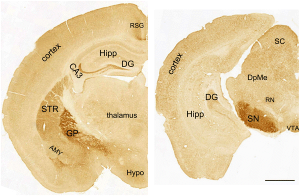
Figure 2 | Mutant Huntingtin Causes a Selective Decrease in the Expression  of Synaptic Vesicle Protein 2C | SpringerLink
Figure 2 | Mutant Huntingtin Causes a Selective Decrease in the Expression of Synaptic Vesicle Protein 2C | SpringerLink

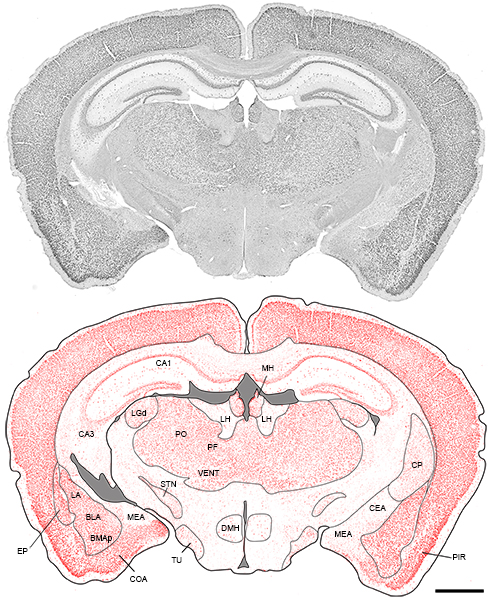
The Mouse Brain Library

File:201510 Mouse brain coronal section.png - Wikimedia Commons

Serial coronal sections of E18.5 mouse brain. Wild type... | Download  Scientific Diagram
Serial coronal sections of E18.5 mouse brain. Wild type... | Download Scientific Diagram

Pictures
Pictures

Molecular atlas of the adult mouse brain

Horizontal (a), coronal (b), and sagittal (c) cross sections from the... |  Download Scientific Diagram
Horizontal (a), coronal (b), and sagittal (c) cross sections from the... | Download Scientific Diagram
Brain, Coronal Section Stock Photo - Alamy

Mouse Brain Atlases
Mouse Brain Atlases

View Image
View Image

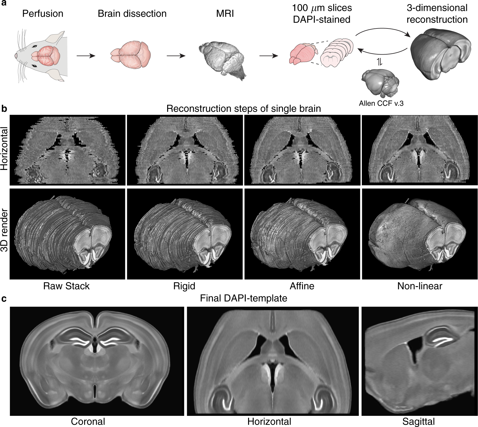
A three-dimensional, population-based average of the C57BL/6 mouse brain  from DAPI-stained coronal slices | Scientific Data
A three-dimensional, population-based average of the C57BL/6 mouse brain from DAPI-stained coronal slices | Scientific Data

High-Throughput Method of Whole-Brain Sectioning, Using the Tape-Transfer  Technique | PLOS ONE
High-Throughput Method of Whole-Brain Sectioning, Using the Tape-Transfer Technique | PLOS ONE

A coronal mouse brain section showing probe placements (illustrated by  vertical lines) in the nucleus of mice used in the present study.
A coronal mouse brain section showing probe placements (illustrated by vertical lines) in the nucleus of mice used in the present study.

Pin on PhD
Pin on PhD

FMRP in Mouse brain | College of Medicine

Mouse Brain, illustration - Stock Image - C030/6193 - Science Photo Library

BioRender | Life Science Icons

BioRender | Life Science Icons

The Mouse Brain Library

Geometry of the coronal and sagittal sections of a mouse brain used... |  Download Scientific Diagram
Geometry of the coronal and sagittal sections of a mouse brain used... | Download Scientific Diagram

File:Coronal section of a mouse brain stained with Hematoxylin & LFB.jpg -  Wikipedia
File:Coronal section of a mouse brain stained with Hematoxylin & LFB.jpg - Wikipedia

A) Schematic representation of a coronal section of mouse brain... |  Download Scientific Diagram
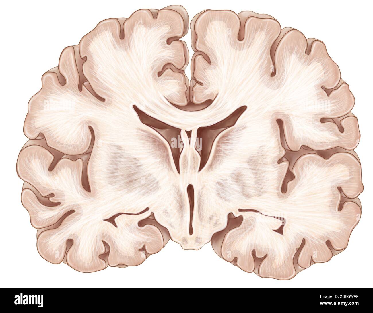
A) Schematic representation of a coronal section of mouse brain... | Download Scientific Diagram
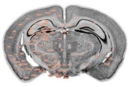
Coronal section of a mouse brain showing the SVZ for harvesting NSCs.... |  Download Scientific Diagram
Coronal section of a mouse brain showing the SVZ for harvesting NSCs.... | Download Scientific Diagram